1. INTRODUCTION
4-hydroxycoumarin, as an important compound of secondary metabolites of the plant kingdom, is a known oral anticoagulant, i.e., the inhibitor of the synthesis of vitamin K-dependent coagulation factors and employed for this purpose for a long time (Anderes and Nand, 2014; Kılıç, 2022). 4-hydroxycoumarin also represents an important compound of various synthetic and natural products with several biological activities, such as anticoagulant, insecticidal, anthelminthic, anti-inflammatory, hypnotic, antifungal, phytoalexin, and HIV protease inhibition effects (Kirkiacharian et al., 2008; Kontogiorgis and Hadjipavlou-Litina, 2005; Lee et al., 2006; Li et al., 2014). These specific advantages of 4-hydroxycoumarin have stimulated its considerable interest in the scientific field, and different 4-hydroxycoumarin derivatives have been synthesized (Kontogiorgis and Hadjipavlou-Litina, 2005; Lee et al., 2006; Li et al., 2014). However, since 4-hydroxycoumarin and its derivatives are soluble in organic solvents and practically insoluble in water, their bioavailability is very low. Additionally, the mentioned compounds are particularly susceptible to interactions with other drugs that can compete with them for plasma protein binding, altering their metabolism in the liver and excretion through the kidneys, resulting in the inhibition or stimulation of the synthesis of the clotting factors that can cause the complications of the diseases (Polifka and Habermann, 2015). With the aim to overcome the mentioned disadvantages, providing controlled release of the bioactive compounds, as well as increasing their effective concentration in the target place, 4-hydroxycoumarin and its derivatives can be encapsulated into numerous carriers.
Liposomal particles are widely employed as carriers for delivering bioactive and non-active compounds, such as proteins, enzymes, polyphenols, vitamins, aromas, and antioxidants in food, pharmaceutical, and cosmetic formulations (Jovanović et al., 2019; Reza Mozafari et al., 2008; Taylor et al., 2005). Liposomes, as biocompatible micro- or nano-sized lipid vesicles, are characterized by one or more phospholipid bilayers, and compared to lipid monolayers, liposomal particles show better fluidity and mobility through the native plasma membrane due to three-dimensional and spherical structure. The main advantages of liposomes, among other encapsulation procedures, are their ability to encapsulate hydrophilic, amphiphilic, and lipophilic compounds, non-toxicity, and biodegradability (Desai and Jin Park, 2005; Jovanović et al., 2019). The liposomal bilayer also provides higher bioavailability of drugs, proteins, nutraceuticals, and polyphenols (Jash et al., 2021; Lee, 2020; Shade, 2016).
Therefore, in the present study, 4-hydroxycoumarin-loaded liposomes were developed and characterized in terms of encapsulation efficiency, vesicle size, polydispersity index (PDI), zeta potential, conductivity, mobility, density, surface tension, viscosity, and 60-day storage stability, as well as antioxidant capacity.
2. MATERIALS AND METHODS
2.1. Plant material and reagents
Distilled water was purified through a Simplicity UV®water purification system (Merck Millipore, Merck KGaA, Germany). 4-hydroxycoumarin, dimethyl sulfoxide (DMSO), 2,2’-azino-bis(3-ethylbenzothiazoline-6-sulphonic acid) or ABTS, copper(II)-chloride, ammonium acetate, neocuproine, methanol, 6-hydroxy-2,5,7,8-tetramethylchroman-2-carboxylic acid or Trolox, and 2,2-diphenyl-1-picrylhydrazyl or DPPH (Sigma-Aldrich, Germany), Phospholipon 90 G (unsaturated diacyl-phosphatidylcholine) (Lipoid GmbH, Germany), ethanol (Fisher Science, UK), and potassium persulfate (Centrohem, Serbia).
2.2. Liposomal preparation
4-hydroxycoumarin-loaded liposomes were prepared using the proliposome method (Jovanović et al., 2022). Phospholipon (2 g), 4-hydroxycoumarin solution in DMSO (66.67 mg/mL, 3 mL), and 5 mL of ethanol were stirred at 50 ◦C. After cooling to room temperature, ultrapure water (15 mL) was added in small portions; the mixture was stirred at 800 rpm for 2 h. The samples were stored at 4 ◦C.
2.3. Determination of encapsulation efficiency
Encapsulation efficiency (EE) of 4-hydroxycoumarin in liposomes was determined using the indirect method. EE was calculated as shown in Equation (1): (1) where miis the initial content of 4-hydroxycoumarin in DMSO solution (66.67 mg/mL) used for the preparation of liposomes, and msupis the content of 4-hydroxycoumarin determined in the supernatant using direct UV-Vis spectroscopy. Non-encapsulated 4-hydroxycoumarin was removed from liposome dispersions by centrifugation at 17,500 rpm and 4◦C for 45 min in Thermo Scientific Sorval WX Ultra series ultracentrifuge (ThermoScientific, USA).
2.4. Antioxidant potential of 4-hydroxycoumarin-loaded liposomes
2.4.1. ABTS method
The ABTS was performed in the following way (Re et al., 1999): (1) ABTS solution (5 mL) and potassium persulfate solution (88 μL) were mixed and left to react for 16 h at 4 ◦C, (2) The obtained mixture was diluted with ethanol (an absorbance of ~0.700 at 734 nm), and (3) ABTS•+solution (2 mL) was mixed with 20 μL of 4-hydroxycoumarin-loaded liposomes. The absorbance was measured after 6 min of incubation, and the ABTS radical scavenging potential (as a percentage of neutralization) of 4-hydroxycoumarin-loaded liposomes was calculated using the following Equation (2): (2) where Axwas the absorbance of ABTS•+solution and the sample (the liposomes or 4-hydroxycoumarin solution), while A0was the absorbance of ABTS•+solution. The ABTS antioxidant potential of 4-hydroxycoumarin solution in DMSO (11.25 mg/mL, the same concentration as in the liposomal sample) was measured as well.
2.4.3. DPPH method
The DPPH radical scavenging potential of 4-hydroxycoumarin-loaded liposomes was determined using the DPPH antioxidant method (Batinić et al., 2022). The liposomal suspension (200 μL) was added in 2.8 mL of ethanol DPPH•radical solution (an absorbance of ~0.800 at 517 nm). After 20 min of incubation, the absorbance was read and the percentage of the inhibition of DPPH• radicals was calculated using the following Equation (3): (3) where Axwas the absorbance of the DPPH•solution and the sample (the liposomes or 4-hydroxycoumarin solution), while A0was the absorbance of the DPPH•radical solution. The DPPH antioxidant capacity of 4-hydroxycoumarin solution in DMSO (11.25 mg/mL, the same concentration as in the liposomes) was also measured.
2.4.3. CUPRAC method
The cupric ion-reducing antioxidant capacity was measured according to the assay described by (Apak et al., 2009). The solution of cupric (II) ion (10−2 mol/mL) was prepared by dissolving 0.0853 g of the copper (II)-chloride dihydrate into 250 mL of distilled water. Ammonium-acetate buffer solution (1 mol/mL) was prepared by dissolving 19.27 g of ammonium acetate in 250 mL of distilled water. The fresh solution of neocuproine was prepared by dissolving 0.078 g of neocuproine in 50 mL of methanol (7.5 × 10⁻³ mol/mL). Each reaction solution consisted of 0.8 mL of 4-hydroxycoumarin-loaded liposomes, 1 mL of copper (II)-chloride solution, 1.2 mL of ammonium-acetate buffer solution, and 1 mL of neocuproine solution. The absorbance was measured at 450 nm after incubation for 30 min in the dark place. Trolox was used as a standard for the calibration curve. The results were expressed as mmol of Trolox equivalents per L (mmol TE/L). Additionally, the cupric ion-reducing antioxidant capacity of 4-hydroxycoumarin solution in DMSO (11.25 mg/mL, the same concentration as in the liposomes) was determined.
All absorbance readings are performed using the UV Spectrophotometer UV-1800 (Shimadzu, Japan). Every spectrophotometric measurement was done in triplicate.
2.5. Photon correlation spectroscopy
The size, PDI, zeta potential, conductivity, and mobility of 4-hydroxycoumarin-loaded liposomes were determined via photon correlation spectroscopy (PCS) in Zetasizer Nano Series, Nano ZS (Malvern Instruments Ltd., UK). The sample was 500-fold diluted and measured three times at room temperature. Additionally, the stability study,i.e., the measurement of the mentioned parameters was repeated on the 30thand 60th days.
2.6. Density, surface tension, and viscosity analyses
The density and surface tension of 4-hydroxycoumarin-loaded liposomes were determined using silicon crystal as the immersion body and Wilhelmy plate, respectively, in Force Tensiometer K20 (Kruss, Hamburg, Germany). Each sample (20 mL) was examined three times at 25 ◦C.
The viscosity of 4-hydroxycoumarin-loaded liposomes was examined using Rotavisc lo-vi device equipment with VOL-C-RTD chamber, VOLS-1 adapter, and spindle (IKA, Germany). Each sample (6.7 mL) was examined three times at 25 ◦C.
2.7. Statistical analysis
The statistical analysis was performed by using the analysis of variance (one-way ANOVA) followed by Duncan’s post hoc test, within the statistical software STATISTICA 7.0. The differences were considered statistically significant at p<0.05, n=3.
3. RESULTS AND DISCUSSION
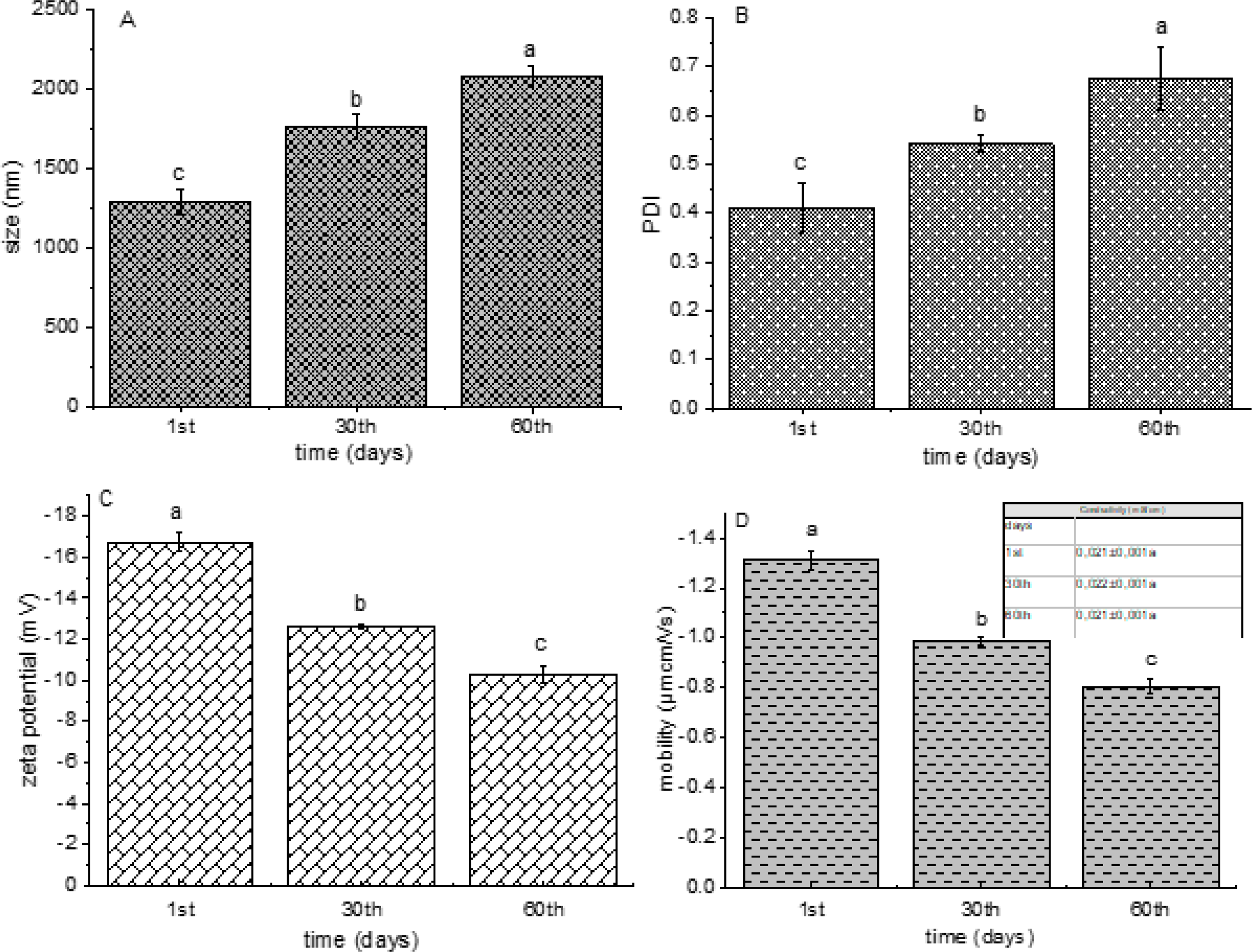
In the present research, 4-hydroxycoumarin-loaded liposomes were developed employing the proliposome technique. Encapsulation efficiency, antioxidant capacity, particle size, PDI, zeta potential, conductivity, mobility, density, surface tension, and viscosity of prepared liposomes were determined. In addition, a 60-day stability study was performed. The results of encapsulation efficiency, antioxidant capacity, density, surface tension, and viscosity are presented in Table 1, while the data from the stability study are shown graphically in Figure 1.
| Sample | EE (%) | ABTS activity (%) | DPPH activity (%) | CUPRAC (mmol TE/L) | density (g/mL) | surface tension (mN/m) | viscosity (mPa•s) |
|---|---|---|---|---|---|---|---|
| liposomes | 96.7±1.2 | 89.76±0.56a | 93.18±0.23a | 0.367±0.003a | 1.007±0.002 | 22.7±0.2 | 14.3±0.2 |
| solution | n.a.* | 90.17±0.10a | 89.82±0.92b | 0.374±0.005a | / | / | / |
In order to determine the efficiency of 4-hydroxycoumarin encapsulation into phospholipid liposomes, the concentration of 4-hydroxycoumarin in supernatant was quantified using direct spectrophotometric analysis. The liposomes were able to encapsulate 4-hydroxycoumarin in a very high yield (96.7%). Our results are in agreement with the literature data (Batinić et al., 2020; Jovanović et al., 2019).
The antioxidant capacity of 4-hydroxycoumarin-loaded liposomes and 4-hydroxycoumarin solution (the same concentration as in the case of the liposomes) was measured using three antioxidant assays, ABTS, DPPH, and CUPRAC methods.
As can be seen from Table 1, the ABTS radical neutralization capacity of the liposomes was 89.76 ± 0.56%, while this value was 90.17 ± 0.10% for the solution of 4-hydroxycoumarin. Therefore, there was no statistically significant difference between the antioxidant capacity of liposomes and solution. The DPPH antioxidant potential of the liposomes and solution was 93.18 ± 0.23% and 89.82 ± 0.92%, respectively (Table 1). It can be noticed that 4-hydroxycoumarin-loaded liposomes showed significantly better antioxidant capacity in the neutralization of free DPPH radicals in comparison to the solution, probably due to the addition of the synthetic antioxidants in the commercial mixture of phospholipids employed for the liposome preparation. Additionally, there was no statistically significant difference between the cupric ion-reducing antioxidant capacity of the liposomes and solution with 4-hydroxycoumarin (0.367 ± 0.003 mmol TE/L and 0.374 ± 0.005 mmol TE/L, respectively, Table 1). The antioxidant potential of natural coumarins and their derivatives has been the subject of intense study for at least two decades and proven in several tests (Ozalp et al., 2020; Todorov et al., 2023). The presented results showed that 4-hydroxycoumarin, although encapsulated into phospholipid liposomal particles retains its antioxidant properties.
Physical characteristics of 4-hydroxycoumarin-loaded liposomes, such as density, surface tension, and viscosity were also measured, and the results are presented in Table 1. The density was 1.007 ± 0.002 g/cm3, surface tension was 22.7 ± 0.2 mN/m, and viscosity was 14.3 ± 0.2 mPa·s. The obtained values of the density and surface tension are in accordance with the data obtained for the liposomes with encapsulated compounds of plant origin (Jovanović et al., 2023). Additionally, (Jovanović et al., 2022) study has reported that the liposomes with plant-origin compounds showed a viscosity of approximately 14 mPa·s.
As can be seen from Figure 1A, the particle size of 4-hydroxycoumarin-loaded liposomes was 1286.3 ± 73.5 nm on the 1st day. The obtained value is in agreement with the literature data, where phospholipid liposomes with encapsulated resveratrol or silibinin (the compounds of plant origin) possessed similar diameter (Isailović et al., 2013; Maheshwari et al., 2011). According to the literature, different factors, such as lipid type, the method for the liposomal formulation, and the physicochemical properties of the encapsulated components significantly influenced the vesicle size of the liposomes (Isailović et al., 2013; Jovanović et al., 2019). The measurements of the vesicle size were repeated on the 30th and 60th days and it can be concluded that there were significant changes in the diameter (1758.0 ± 77.6 nm and 2077.3 ± 63.2 nm, respectively, Figure 1A). The increase in the size was expected since the sample showed a relatively low value of zeta potential, particularly after the 30th and 60th days (described below, Figure 1C).
PDI values, as a measure of the particle size distribution in the suspension, were determined in 4-hydroxycoumarin-loaded liposomes suspension on the 1st, 30th, and 60th days as well, and the data are shown in Figure 1B. PDI value was 0.409 ± 0.050 on the 1st day indicating the existence of a moderately dispersed distribution (Ardani et al., 2017). The method employed for the preparation of the liposomes significantly influences the uniformity of the liposomal population (Isailović et al., 2013; Jovanović et al., 2019). PDI values significantly increased during the 60-day storage study, as in the case of liposome diameter (0.542 ± 0.019 on the 30th day and 0.676 ± 0.064 on the 60th day, Figure 1B).
The zeta potential, as a measure of the stability of 4-hydroxycoumarin-loaded liposomes, was monitored during 60 days of storage at 4 ◦C and the results are presented in Figure 1C. The zeta potential was -16.73 ± 0.47 mV on the 1st day, which indicates the presence of a moderately stable liposomal system. Phosphatidylcholines, as neutral lipids in the water surrounding, can be reoriented causing the presence of a surface charge, i.e., zeta potential. Namely, the negative value of zeta potential is related to the exposure of the phosphate group lying in an outer plane concerning the choline groups (Jovanović et al., 2019). However, after the 30th and 60th days, a statistically significant drop can be noticed in the absolute values of zeta potential (-12.63 ± 0.06 mV and -10.31 ± 0.42 mV, respectively, Figure 1C). Therefore, the results of zeta potential prove that liposomal vesicles with 4-hydroxycoumarin were not stable during the 60 days of storage at 4 ◦C. The absence of stability was also confirmed by the significant changes in the particle size and PDI values (Figures 1A and 1B), i.e., fusion or fission of the liposomes probably occurred.
The conductivity of the liposomal suspension is affected by the exposed charge of the phospholipid components and correlates to a volume of liposome entrapment. The measured conductivity of 4-hydroxycoumarin-loaded liposomes was 0.021 ± 0.001 mS/cm on the 1st day (table in Figure 1D), as in the case of plain phospholipid liposomes described in a previous study (Jovanović et al., 2022). Considering that there were no changes in the conductivity values of the liposomes with 4-hydroxycoumarin during the 60 days of storage, it can be concluded that there was no leakage of the encapsulated compound from the liposomal vesicles. Hence, the increase in conductivity values during the storage of liposomes is usually related to the leakage of the encapsulated components. The mobility of liposomal particles represents a function of size, zeta potential, and lipid composition (Duffy et al., 2001). The mentioned parameter was measured on the 1st, 30th, and 60th days and the results are shown in the graph of Figure 1D. The mobility significantly decreased during storage (from -1.31 ± 0.03 μmcm/Vs and -0.806 ± 0.03 μmcm/Vs), as in the case of zeta potential. The decrease in mobility can be explained by the increase in vesicle size.
4. CONCLUSION
In the present research, 4-hydroxycoumarin-loaded liposomes were developed and characterized in terms of encapsulation efficiency, antioxidant potential, particle size, PDI, zeta potential, conductivity, mobility, density, surface tension, viscosity, and storage stability. The liposomal population showed a very high level of encapsulation capacity, as well as the ABTS and DPPH radical scavenging and cupric ion-reduction antioxidant potential. The values of the density, surface tension, and viscosity meet the criteria for the product that can be further used. However, size, PDI, zeta potential, and mobility significantly changed during the 60-day storage study indicating the existence of an unstable liposomal system. On the other hand, the conductivity of the liposomes with 4-hydroxycoumarin did not vary, thus, it can be concluded that there was no leakage of the encapsulated compound. Therefore, future research should include the optimization of the lipid composition with the aim of improving 4-hydroxycoumarin-loaded liposome stability and investigation of other biological effects in vitro and in vivo.


